
Steps to a Comprehensive Oral and Radiographic Evaluation (CORE)
Go beyond the surface. A complete CORE procedure transforms a routine cleaning into the gold standard of veterinary dental care
By Denise Rollings, AAS, CVT, VTS (Dentistry), Key Account Manager, iM3 | 2026
Veterinary dentistry is not just about cleaning teeth or eliminating bad breath. Periodontal disease is the most common condition diagnosed in both dogs and cats, with 80% of pets over the age of three affected. Dental procedures are an important component of well-rounded veterinary care and should be treated with the same priority and precision as other medical procedures.
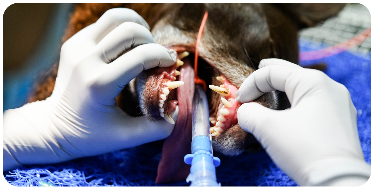
Dental procedures are performed under anesthesia and require skilled, trained veterinarians and veterinary technicians. Only veterinarians can diagnose disease, but veterinary technicians play a critical role in supporting diagnosis and treatment. Technicians assist by obtaining accurate histories, recognizing pathology, and communicating findings to the veterinarian. They also perform initial workups, periodontal probing, charting, scaling, polishing, taking diagnostic radiographs, and administering nerve blocks—demonstrating just how central their role is in these procedures.
Technicians can be trained in all aspects of a thorough dental procedure, including performing complete dental cleanings, capturing diagnostic radiographs, and administering nerve blocks. With the high prevalence of periodontal disease in pets, routine dental care should be part of every patient’s preventive health plan.
AFFECTED BY
PERIODONTAL DISEASE
NOT VISIBLE
TO THE EYE
The Pre-anesthetic Exam:
Begin with an oral history and visual examination. Ask pet owners about signs such as bad breath, swollen or bleeding gums, drooling, changes in eating or chewing habits (e.g., dropping food, approaching food but not eating, vocalizing during meals), tooth loss, and reduced interest in toys.
Pets often mask signs of pain or discomfort, making owner input essential. Ask what the pet prefers to chew on and whether any home dental care is currently being provided. Pain-related aversion to toys or foods is often misinterpreted as behavioral preference.
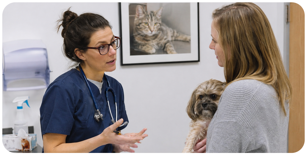
Clinical signs of oral disease observed on a conscious exam may include halitosis, malocclusions, persistent deciduous teeth, plaque and calculus buildup, missing teeth, and signs of periodontal disease such as gingival recession or tooth mobility.
Running blood work is essential. While age is not a disease, older pets are more likely to have concurrent health issues. Ideally, diagnostics should be performed prior to the procedure day to avoid last-minute cancellations and allow time for protocol adjustments. Recommended testing may include a complete blood count, serum chemistry panel, urinalysis, coagulation panel (if indicated), chest radiographs, echocardiogram, ECG, and blood pressure measurement.
Protecting yourself: Personal Protection Equipment (PPE)
Wear eye protection. Goggles, loupes, or a visor should be worn by everyone in the operatory area as pieces of calculus, tooth, bone, blood, saliva, and even broken burs become airborne and can cause an eye injury or infection. Surgical face masks that cover the mouth and nose should be worn.
Anti-fog masks are helpful in preventing protective eyewear from fogging up. Appropriately fitted gloves and protective clothing such as a gown or lab coat, as well as surgical caps should be worn to prevent bacterial contamination of clothing and hair. You should also consider hearing protection if the noise of the dental unit running is bothersome.
Ergonomics is vital to preventing fatigue and injury. Dentistry has a lot of small and repetitive movements that can strain our muscles, tendons, and ligaments. To protect ourselves from fatigue and short- or long-term injury, we should practice the best ergonomics possible.
Adjust the table to fit the individual, allowing arms to be parallel with the table. A dental saddle seat allows one to sit with a straight back, their thighs slightly angled to the floor, and their gaze looking down without bending over. Utilizing a modified pen grasp when holding instruments reduces strain of repetitive movements. Fatigue mats can be used if one is standing for prolonged periods of time.
Complete Oral Exam and Charting
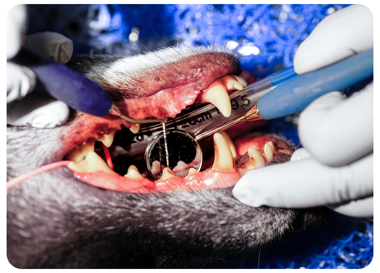
The complete oral exam should be performed in a systematic manner with the patient under general anesthesia. Our goal is to find pathology, treat what needs to be treated, and prevent disease.
We need to shift our emphasis from cleaning the teeth when they look dirty or extracting teeth when they are falling out, to prevent disease from getting that bad in the first place. The oral exam is as or more important than the cleaning.
Periodontal Cleaning:
After the complete oral exam is completed, the dental cleaning is performed.
Supragingival Plaque and Calculus Removal
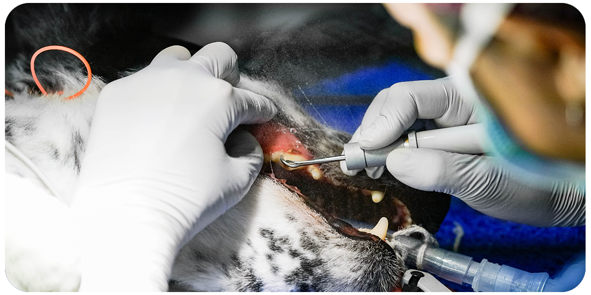
Remove supragingival plaque and calculus. This is most important to the owner because it is what they see, however it is the least important to the patient. Use the powered (ultrasonic, magneto restrictive, or piezo) scaler to begin with. Do not spend more than ten seconds on a tooth at a time and be sure you are using enough water to cool and irrigate the tooth as you work.
Subgingival Cleaning
Remove subgingival plaque and calculus with a curette or perio tip. This is the least visible to the owner but the most important to the patient. Plaque bacteria are the cause of periodontal disease, not calculus. The plaque must be removed from any subgingival space. Plaque is a biofilm that contains bacteria.
What is Plaque?
Plaque is the filmy, soft deposit on our teeth that can be removed by brushing. Plaque then mineralizes to form calculus, which is also referred to as tartar. The minerals come from saliva. The hard, rough calculus increases the surface area for more plaque to stick to.
The biproducts of the plaque bacteria are toxic to gingival tissues. They can break down collagen along with the hosts’ immune response and bone, eventually leading to attachment loss and destruction of the periodontium. Contributing factors to periodontal disease are malocclusions including crowded and rotated teeth, calculus, restorations, orthodontics, genetics, xerostomia, gingival enlargements, and systemic health.
Always Check Your Work
Use the air water syringe to rinse the mouth of calculus and blood, as well as drying the teeth when you are finished. Look for the chalky white residue that remains on the teeth. That is the calculus that has been missed. Remove what is left with a hand scale and/or curette.
Polishing
Polish the teeth using fine or flour grade paste above and below the gum line. Flare the polishing cup under the gingival sulcus. Use a light touch and spend five to ten seconds or less per tooth. Polishing smooths the tooth surface and removes irregularities created by scaling. This decreases the surface area for plaque bacteria to stick to. Once all the calculus and plaque have been removed and teeth polished, rinse the tooth surface and sulcus to remove debris and paste.
Applying Fluoride
At this point, fluoride may be applied, which is an ingredient used to decrease tooth sensitivity. However, you should not use this in renal patients, as fluoride is excreted through the kidneys and our patients cannot spit it out.
"Obtain full mouth radiographs! Radiographs are necessary and are part of comprehensive oral exams."
Full Mouth Radiographs
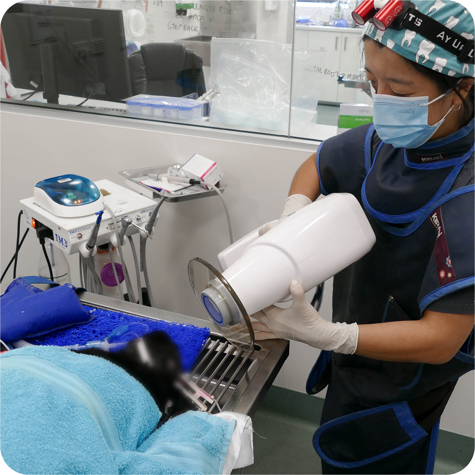
Radiographs are necessary and are part of comprehensive oral exams. We can see the crown and probe to determine periodontal pockets, but we cannot see what is below the gumline. Radiographs tell us if a tooth can be repaired or if it needs to be removed. This is an important diagnostic tool.
Most of the pathology is below the gum line, and 80% of the dental anatomy is not visible. Radiographs must be obtained post extractions as well, to determine the entire tooth is removed and no fragments are left behind.
Treatment Plan
A treatment plan should be customized for each tooth in every patient based on the complete oral exam, probing, and intraoral radiographs. A veterinarian must perform surgery, extractions, and advanced dental procedures.
Take post procedure photographs of both sides but be sure to cover the endotracheal tube with the tongue so it does not look too scary to the owner in the picture.
Discharge Instructions:
Personalize the instructions based on the individual’s condition. Include feeding instructions, medications, and recheck dates. Have the directions and instructions for medications to go home in writing for the owner. It should include when the owner should start the medications, how often to give them, how many to give, what the medication is for, and for how many days.
If oral surgery was performed, the pet needs to be rechecked within seven to14 days post operatively to make sure all sites are healed. They should also come back in three-month intervals for an oral exam. This is decided by not only the extent of periodontal disease present, but also the breed of your patient.

Toy breeds are more predisposed to periodontal disease than large or giant breed dogs. Discuss this with the owner. Put reminders in the computer to generate a reminder for future appointments.
Discuss home care with the owner when they bring the pet back for the follow up exam. They are usually overwhelmed at the time of surgery and may not understand the conversation.
Send a packet home with samples of all the products you carry that may be appropriate for that patient. Explain what they are and how they work. Now that the teeth are nice and clean, we need to keep them that way. Bacteria are already multiplying as they are walking out the door.
Educate the client on the importance of home care to help protect their investment. Proper dentistry takes a team, but it can be achieved so we can improve the lives of pets, one tooth at a time.
